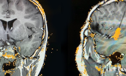
A new study reports obliteration rates above 80% for treating arteriovenous malformations (AVMs) with single-fraction stereotactic radiosurgery (SRS), also known as Gamma Knife Radiofrequency (GKRS). The study, recently published in Child’s Nervous System, spans 17 years of patient care at Children’s HealthSM.
The cumulative incidence of obliteration was 83% at 5 years for all patients (n=89) and 90% among patients followed for 3 years or more (n=59). Standard rates reported elsewhere range from 64% to 77%.
The study represents one of the largest single-center patient series in radiosurgery for pediatric AVMs and underlines why the treatment approach was effective. That approach includes 3 distinct strategies:
Avoiding embolization prior to radiosurgery
Recommending radiosurgery to many patients with low Spetzler-Martin (S-M) Grading Scale scores
Strategic use of MRI and angiography during surgery planning and postoperative follow-up
“Other centers that adopt these practices may improve their radiosurgery outcomes and help more children avoid recurring AVMs, neurological deficits and other associated problems,” says Bradley Weprin, M.D., Division Chief of Neurosurgery at Children’s Health and Professor at UT Southwestern.
High obliteration rate with only temporary complications
The retrospective study included patients receiving care at Children’s Medical Center Dallas from 2006–2023. All patients received stereotactic radiosurgery in a single session, and 90% of AVMs (91/101) were treated with 18–20 Gy to the 45–60% isodose line.
The obliteration rate increased over time, with a cumulative incidence of 55.0% at 3 years, 73.1% at 4 years, and 83.0% at 5 years in the full patient cohort. The median time to obliteration was 31 months.
No hemorrhages or secondary malignancies occurred. Many patients experienced temporary headaches and residual neurological deficits. Most of these patients had a history of prior hemorrhage or AVM treatment, and all symptoms resolved over time.
Only 8.5% experienced symptomatic radiation-induced change, also presenting primarily as headaches, neurologic deficits or seizures. Once again, all cases were temporary.
“When a patient developed new or worsening seizures, they went away. When patients experienced a neurological deficit, it didn’t last,” says Dr. Weprin, who also serves as Board President of the American Society of Neurosurgeons.
Avoiding embolization prior to radiosurgery
Some patients in the study (13.5%) received embolization at another center prior to treatment at Children’s Health. They required radiosurgery because large embolized vessels recanalize eventually.
“We embolize some vessels prior to open surgery, but not prior to radiosurgery,” says Dr. Weprin. “It takes 2–3 years for obliteration to occur after radiosurgery, and the embolized vessels can reopen in that time.”
Endovascular surgeons are now able to embolize the smaller vessels within an AVM, and those vessels do not reopen. But the risk of complications is higher, so the Children’s Health team takes this route only when they’re confident it’s safe.
“We discuss every patient and agree on the right option: open surgery, radiosurgery or microsurgery,” says Dr. Weprin.
Offering radiosurgery to patients usually reserved for open surgery
The study included many patients at level 1 (12.9%) and 2 (39.6%) on the Spetzler-Martin scale, who typically receive open surgery. The high obliteration rate and low toxicity observed in the study show that these patients are often good candidates for radiosurgery.
In cases where the team determines that open surgery and radiosurgery are both viable, the right option depends on the patient’s risk tolerance and lifestyle priorities. Open surgery has the advantage of immediate obliteration, whereas radiosurgery comes with a latency period.
“But radiosurgery is a day surgery with fewer activity restrictions, no scars and no risk of residual AVM from incomplete resection,” says Dr. Weprin. “We talk through the short- and long-term tradeoffs and help patients reach a decision they feel confident about.”
Using MRI and angiography together
The team plans surgery by performing an MRI and angiogram simultaneously and merging the images. Some centers use MRI alone, but AVMs can evade detection.
In fact, Dr. Weprin once did an internal experiment in which he, a neuroradiologist and an adult neurosurgeon each circled the AVMs they saw on MRI and compared their results to an angiogram. “We were all off,” he says.
Angiography is the gold standard, and combining it with MRI allows the team to “round the corners” and see tissue more fully. All images are interpreted by a pediatric neuroradiologist rather than a general practitioner.
The team also uses both tests during follow-up, but in series: Patients get an MRI every 6 months after radiosurgery, and an angiogram at 3 years – or sooner, if an MRI suggests obliteration.
“Obliteration often occurs more quickly in children than in adults,” says Dr. Weprin. “We want to confirm it as soon as possible, so kids can stop wondering and move on with their lives.”
Learn more about innovations in pediatric neurosurgery at Children's Health.